排泄器官查看源代码讨论查看历史
 |
排泄器官是将机体新陈代谢过程中产生的终产物排出体外的器官。 由于动物进化水平不同和适应多变环境的结果,排泄器官一般可分为下列5类:①原生动物的伸缩泡。②昆虫的马尔皮基氏小管。③甲壳动物的触角腺。④无脊椎动物的肾器官,包括原肾管、后肾管和肾管。⑤脊椎动物的肾脏。
脊椎动物
脊椎动物在胚胎时期都有前肾出现,但只有鱼类和两栖类的胚胎时期,前肾才有作用。圆口纲中的盲鳗仍用此种肾脏作为排泄器官。
前肾
前肾的位置靠近体腔的前段,由许多排泄小管(肾小管renal tubules)组成。小管的一端开口于体腔,开口处膨大成漏斗状,其上着生纤毛,这就是肾口,可以直接从体腔内收集代谢废物。在肾口附近还有由血管丛形成的血管球,它们利用滤过血液的方式把血中的废物排出送入排泄小管中。小管的另一端与一个总的管道相通。这个总管称为前肾导管,其末端通到体外。
中肾
中肾是鱼类和两栖类成体阶段执行排泄机能的器官,位于前肾的后方。排泄小管的肾口已显出退化,有些小管的肾口甚至完全退化,不能直接与体腔相通。靠近肾口附近的排泄小管外凸成为小支,小支末端膨大内陷成为双层的囊状结构,叫做肾球囊(包曼氏囊),把血管球包入其中,共同形成肾小体(malpighian body)。肾小体和它的排泄小管一起组成一个泌尿机能的基本构造,特称为肾单位(nephron)。
在中肾阶段,原来的前肾导管纵裂为二,其一成为汇集排泄小管尿液的总管道,即中肾导管,在雄性动物兼有输精管的作用;另一管在雄体已退化,在雌体则演变为输卵管。
后肾
后肾是爬行类、鸟类、哺乳类成体的排泄器官,其位置靠近体腔的后段。外部形态因动物种类不同而有差别。后肾的排泄小管末端只有肾小体,肾口已完全消失。各肾小管运送尿液汇入的总管道,就是后肾导管(前肾导管,中肾导管和后肾导管,通常亦泛称为输尿管)。此管是由中肾导管生出来的一对突起,向前延伸成管,各和一个后肾连接。后肾发生后,中肾和中肾导管都失去了泌尿的机能而转成生殖系统的组成部分,中肾导管完全成为输精管,遗留下来的中肾排泄小管则形成副睾等构造。
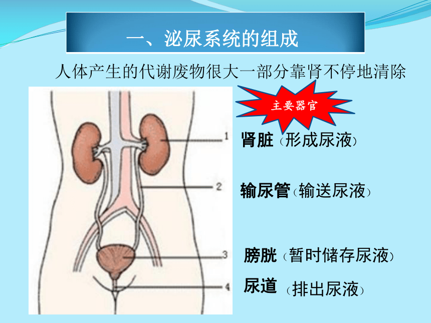
后肾是脊椎动物肾脏中最高级的类型。就哺乳类而言,它的后肾是一对常呈豆形的主要的排泄器官,滤出尿液,经输尿管,膀胱和尿道而排出体外。
排泄作用是指生物体将代谢废物排出体外的作用,是所有生物生存的必要过程。单细胞生物透过细胞表面排出废物。高级植物以叶面上的气孔排气。多细胞生物则有特别的排泄器官。其中有参与排泄作用的器官(其他系统的一部分),也可归类到排泄系统(Excretory system)的一部分。
无脊椎动物
(一)无特殊排泄器.如腔肠动物等,由各个细胞直接进行气体交换和排出废物;棘皮动物由体腔液中的变形细胞收集代谢产物,穿过皮鳃逸出体外。
(二)收缩泡.见于淡水产原生动物及多孔动物.
(三)原肾型.见于扁形动物、有假体腔的动物及环节动物和软体动物的幼虫.基本构造为1对纵行的排泄管及数目不等的焰细胞,焰细胞中空,内有1束不断摆动的纤毛,把收集的废物送至排泄管,由排泄孔排出.但线虫类只有1个由原肾细胞衍生的管状或H状细胞,细胞内无纤毛,排泄管为细胞内管道.
(四)后肾型.见于环节动物和软体动物.基本构造包括开口于体腔的1对或多对腺状器官,由肾孔排出代谢废物.环节动物的后肾管(外胚层来源)可和中胚层来源的体腔导管合成共同的管道及排泄孔.有的种类也可由原肾管与体腔导管合成.有些甲壳纲动物具有类似后肾管的排泄器,称[[触角腺.
(五)马氏管.见于陆栖的节肢动物.马氏管呈丝状,自2条至数百条,浸于血液中.马氏管的管壁只有1层细胞,末端为盲端,另一端通入肠道,代谢废物由肛门排出体外.
英文翻译:Invertebrates to rid the body of waste organs. The general sub-situations: (1) no special drainage devices. Such as coelenterate, etc. directly from the various cells, gas exchange and discharge of waste; echinoderms from the deformation of body cavity fluid cell collection metabolites across the skin gills escape in vitro. (2) contraction bubble. protozoa found in fresh water and porous animal production. (3) of the original kidney. found in Platyhelminthes, there are fake body cavity of animals and part of animals and molluscs larvae. the basic structure for the 1 on the longitudinal excretory duct and the number of lines, ranging from flame cell flame cell hollow, containing a bundle of cilia continue to swing to the collection of waste sent to the excretory duct, excretory pore from the exhaust. But the line of insects is only one original cell-derived renal tubular or H-shaped cells, nonciliated cells, excretory duct cells of pipes for. (4) after the kidney-type. found in annelid and molluscs. the basic structure, including openings in the body cavity of the 1 on one or more pairs of glandular organs, metabolic wastes discharged by the kidneys Kong. annelid post-renal tube (ectodermal origin) and the mesoderm may be the source of the body cavity tube synthesis of the common ducts and excretory pore. Some species can also be the original kidney tube and body cavity catheter synthesis. some crustacean similar excretion of renal tube device, saying antennary gland. (5) Malpighian tubules. found in terrestrial arthropods. Malpighian tubules was filamentous, from two to hundreds of Article immersed in the blood. Malpighian tubules of the wall is only one layer of cells, the end of the blind side, other side of the pass into the gastro-intestinal, metabolic wastes excreted by the anus.